


Content of
Digestive System Physiology & Anatomy PowerPoint Presentation
Slide 1: Digestive System Anatomy and Physiology Cover Slide

This presentation provides an overview of the anatomy and physiology of the digestive system.
Slide 2: The GI Tract Anatomy Function and Role in Digestion

The gastrointestinal tract is a muscular tube that extends through the abdomen. It serves three primary functions. the mechanical and chemical breakdown of food. the absorption of nutrients into the bloodstream. and the elimination of waste through defecation.
Slide 3: The GI Tract Wall Structure and Layers

The layers of the gastrointestinal tract wall, include the following. the mucosa, which consists of an epithelial lining, responsible for nutrient absorption and mucus secretion. the submucosa, a layer of connective tissue containing blood vessels, nerves, and glands. the muscularis propria, composed of smooth muscle layers that facilitate peristalsis and the movement of food. and the serosa, an outer protective layer that minimizes friction with surrounding tissues.
Slide 4: Enteric Muscular System Structure and Function

The enteric muscular system is composed of several layers, each with distinct structural and functional characteristics. These include. the epithelium, which forms a protective barrier and facilitates nutrient absorption. the longitudinal and circular muscle layers, responsible for peristalsis and segmentation. the submucosa, a supportive layer housing blood vessels, nerves, and lymphatics. finally the muscularis mucosae, a thin layer of smooth muscle that aids in local movement of the mucosa to enhance secretion and absorption.
Slide 5: The Mucosa Layer Structure and Function

The mucosa layer, the innermost layer of the gastrointestinal tract, plays a pivotal role in nutrient absorption and immune defense. This layer is composed of three distinct sublayers:
First, the Epithelium: This surface layer is directly responsible for absorption and secretion processes, facilitating the exchange of nutrients.
Second, the Lamina Propria: A connective tissue layer containing blood vessels, nerves, and immune cells.
Finally, the Muscularis Mucosae: This thin muscle layer enhances the mucosal surface by inducing gentle movements, promoting efficient absorption and secretion along the GI tract.
Slide 6: The Submucosa Layer Structure and Functions

The submucosal layer, which serves as the structural support framework within the gastrointestinal tract contains. a rich network of blood vessels that supply essential nutrients and oxygen to surrounding tissues. Additionally, this layer houses intricate nerve plexuses responsible for coordinating smooth muscle contractions and secretions. Finally, the submucosa includes lymphatic vessels that contribute to fluid drainage and transport immune cells.
Slide 7: The Muscularis Propria Layer Structure and Function

The muscularis propria is a critical layer of the gastrointestinal tract, responsible for propelling food through peristaltic movements. This layer consists of two distinct muscle layers: the inner circular muscle layer. which contracts radially to narrow the lumen. and the outer longitudinal muscle layer. which contracts along the length of the tract to shorten and propel contents forward.
Slide 8: The Serosa and Adventitia Layers Structure and Functions

the serosa or adventitia layer, is The outermost layer. it varies in structure and location based on the organ's position within the body. Specifically, serosa is present in organs within the peritoneal cavity, for example; the liver, the stomach, and the small intestine. On the other hand, the adventitia, a connective tissue layer, surrounds retroperitoneal organs, for example; the kidneys, the pancreas and some parts of the large intestine.
Slide 9: Epithelial Diversity in the GI Tract and Its Functions

The gastrointestinal tract exhibits notable epithelial diversity, reflecting its specialized functions across different regions. In the esophagus, stratified squamous epithelium provides a protective layer against abrasion during the passage of food. Moving into the stomach and small intestine, the simple columnar epithelium facilitates absorption and secretion processes essential for digestion. Additionally, goblet cells, which are dispersed throughout various sections of the GI tract, play a crucial role in mucous secretion, protecting the mucosal lining and aiding in smooth content passage.
Slide 10: The Peritoneum Structure and Function in Abdominal Cavity

The peritoneum is a serous membrane that lines the abdominal cavity and envelops its organs, facilitating smooth, friction-free movement within the abdominal space. It consists of two main layers. the parietal peritoneum, which adheres to the inner abdominal wall. and the visceral peritoneum, which covers the surfaces of abdominal organs. Between these layers lies the peritoneal cavity, a potential space filled with serous fluid.
Slide 11: Layers of the Peritoneum and Their Functions in Abdominal Anatomy

to explore their anatomy more. The parietal peritoneum lines the abdominal wall, and extends to fuse with the diaphragm, and retroperitoneal organs. anchoring these structures, and contributing to the structural integrity of the abdominal cavity. In contrast. the visceral peritoneum envelops the abdominal organs. and plays a role in forming anatomical structures. that help secure organs in place.
Slide 12: Key Support Structures of the Peritoneum Ligaments, Omenta, and Mesenteries

The folds and support structures of the peritoneum include. the Ligaments, consist of peritoneal folds that connect organs to the abdominal wall or to each other, stabilizing their placement. The omenta are double-layered folds extending from the stomach to other organs, providing both protection and pathways for blood vessels. Finally, the mesenteries are peritoneal extensions that anchor the intestines and other abdominal organs to the posterior abdominal wall, supporting vascular and neural connections essential for organ function .
Slide 13: The Mouth Structure and Functions in the Digestive System

The mouth is a muscular cavity that marks the beginning of the digestive tract. It is bounded by the lips anteriorly, the cheeks laterally, and the hard and soft palate superiorly. The mouth contains several important structures, including the teeth, which are responsible for mechanical digestion; the tongue, which aids in taste, swallowing, and speech; and the salivary glands, which secrete saliva to facilitate digestion and oral hygiene.
Slide 14: Functions of the Tongue in the Digestive Process

Inside the mouth, the tongue serves four primary functions. First, it assists in the mechanical breakdown of food by manipulating and mixing it with saliva, which facilitates digestion. Second, the tongue is essential for taste perception, as it contains taste buds that detect various flavors. Third, it plays a crucial role in speech articulation, helping to form sounds and words. Finally, the tongue is involved in the initiation of swallowing by pushing food to the back of the mouth, thereby beginning the swallowing process. Together, these functions highlight the tongue’s importance in both digestion and communication.
Slide 15: Salivary Glands Anatomy

The salivary glands consist of three major pairs. and located in the mouth. These glands are. the parotid glands, they are the largest, and located near the ears. the submandibular glands, are situated beneath the jaw. and the sublingual glands, are found beneath the tongue. These glands secrete saliva, which aids in digestion by moistening food, facilitating chewing and swallowing.
Slide 16: Anatomy of the Pharynx Nasopharynx, Oropharynx, and Laryngopharynx

The pharynx is divided into three distinct parts: the nasopharynx. is connected to the nasal cavity and serves as a passage for air. the oropharynx, which is continuous with the oral cavity and serves as a common passage for both air and food. and the laryngopharynx, which connects to the larynx and esophagus, directing food and liquids into the esophagus while allowing air to enter the larynx and trachea.
Slide 17: Structure and Layers of the Esophagus

The esophagus consists of four distinct layers, each serving a specific function to facilitate the movement of food from the mouth to the stomach.
The Mucosa: The innermost layer, which provides protection against abrasion from food particles as they pass through the esophagus.
The Submucosa: Located beneath the mucosa, it contains blood vessels, lymphatics, and nerve plexuses, which support the esophagus and contribute to its function.
The Muscularis Propria: This layer consists of smooth muscle responsible for peristalsis. Finally The Adventitia: The outermost layer, made of connective tissue, anchors the esophagus to surrounding structures, ensuring its stability
Slide 18: Anatomy of the Stomach

The stomach is a muscular sac located in the upper left part of the abdomen, playing a central role in digestion. It begins at the cardia, where the esophagus meets the stomach, and is divided into several sections. the fundus, which is the upper portion that stores food. the body, which is the largest section responsible for the mixing of food and gastric juices. the antrum, which prepares the food for passage into the small intestine. and the pylorus, the final section that connects to the duodenum and regulates the flow of partially digested food into the small intestine.
Slide 19: Stomach Histological Features

The histological features of the stomach are crucial to its digestive functions and protection against harsh conditions. The mucus layer, which coats the stomach lining, plays a vital protective role by shielding the mucosal surface. Gastric pits are invaginations in the epithelial lining of the stomach. At the base of these gastric pits, gastric glands contain various specialized cells, that contribute to the overall secretion of digestive enzymes.
Slide 20: Gastric Glands Explained

The gastric glands in the stomach contain several specialized cells that each play a critical role in digestion and the protection of the stomach lining. Surface Mucous Cells produce a thick layer of mucus that acts as a protective barrier. Mucous Neck Cells, found deeper in the gastric pits, secrete additional mucus that enhances this protective layer. Parietal Cells are responsible for secreting hydrochloric acid, which creates the acidic environment necessary for activating digestive enzymes and facilitating the breakdown of food. Enteroendocrine Cells secrete hormones, such as gastrin. Chief Cells secrete pepsinogen, an inactive enzyme precursor, which is activated in the acidic environment to form pepsin. Together, these cells ensure effective digestion while protecting the stomach lining from self-digestion and damage.
Slide 21: Function of the Pyloric Sphincter in Regulating Digestion

The pylorus is found at the end of the stomach. it is the distal portion of the stomach and contains. the pyloric sphincter, a muscular valve that tightly regulates the flow of partially digested food know as the chyme. ensuring the chyme is released in small amounts.
Slide 22: The Pancreas Anatomy

The pancreas is a flattened elongated gland that plays a crucial role in both endocrine and exocrine functions. It is divided into five main regions: the uncinate process, which is a hook-like projection extending from the lower part of the head of the pancreas; the head proper, the broad part that lies within the curve of the duodenum and is significant for digestion; the neck, a narrow region located between the head and body, positioned anterior to the superior mesenteric vessels; the body, which is the central portion extending horizontally across the midline of the abdomen; and finally, the tail, the tapered end that lies near the spleen. Together, these regions enable the pancreas to effectively manage digestion and regulate blood sugar levels.
Slide 23: The Pancreas Exocrine and Endocrine Functions

There are two distinct functional components.
First is the Exocrine Pancreas: This is the larger portion of the pancreas, responsible for producing digestive enzymes. These enzymes are secreted into the duodenum via the pancreatic duct, where they aid in the digestion of food.
Second. the Endocrine Pancreas: Consisting of clusters of cells called the islets of Langerhans, that secrete hormones directly into the bloodstream.
Slide 24: The Pancreas Acinar Cells and Pancreatic Islets Functions

The pancreas consists of several distinct cell types that contribute to its exocrine and endocrine functions. The acinar cells, located in the exocrine portion of the pancreas, produce digestive enzymes, and play a vital role in the breakdown of food in the small intestine. Scattered throughout the pancreas are Islets of Langerhans, clusters of endocrine cells that secrete hormones directly into the bloodstream. Within the islets the following is found. alpha cells: produce glucagon, which raises blood glucose levels. beta cells secrete insulin to lower blood glucose levels, in addition to producing amylin, which helps regulate blood sugar levels postprandially. Delta cells secrete somatostatin, which inhibits the release of both insulin and glucagon. Epsilon cells release ghrelin, a hormone that regulates hunger. PP cells produce pancreatic polypeptide, which regulates pancreatic secretions and appetite control. Together, these cells play a key role in regulating digestion, metabolism, and energy homeostasis.
Slide 25: The Liver Anatomic and Location in the Abdomen

The liver occupies a prime location in the upper right quadrant of the abdomen. It rests beneath the diaphragm, the muscular sheet that separates the chest and abdominal cavities. Protected by the lower ribs, the liver sits superior to the stomach, intestines, and the right kidney.
Slide 26: Anatomy of the Liver Key Structures and Ligaments Overview

In the anterior view of the liver, several key structures are visible. The inferior vena cava. a large vein running along the posterior surface of the liver. responsible for returning deoxygenated blood from the lower body to the heart. The right lobe. the larger of the two main lobes of the liver. Beneath the right lobe lies the gallbladder. a small organ that stores bile produced by the liver. releasing it into the small intestine to aid in digestion.
On the left side of the liver. additional structures are observed. The left lobe. smaller than the right lobe. The falciform ligament. a fold of tissue, attaches the anterior part of the liver to the anterior abdominal wall and diaphragm. providing structural support. Lastly. the teres ligament, a fibrous remnant of the fetal umbilical vein.
Slide 27: Posterior View of the Liver with Key Anatomical Structures

In the posterior view of the liver, several important structures can be identified. The inferior vena cava known as the IVC. runs along the posterior surface. Connected to the IVC are the hepatic veins. which drain deoxygenated blood from the liver into this major Vein.
The caudate lobe. a small lobe situated near the IVC. plays a role in liver function and bile production. The portal vein is also visible. transporting nutrient-rich blood from the gastrointestinal tract and spleen to the liver for processing. Additionally, the common bile duct. which delivers bile from the liver and gallbladder to the duodenum.
Supplying oxygenated blood to the liver is the function of the proper hepatic artery. Finally, the quadrate lobe. located on the liver's inferior surface adjacent to the gallbladder.
Slide 28: Structure and Function of Hepatocytes, Sinusoids, and Bile Canaliculi

the liver’s microscopic structure reveals a highly organized system. designed to support its vital functions. At the core of this system is the lobule. a hexagonal-shaped structure that serves as the liver's fundamental functional unit. Each lobule is centered around a central vein that collects blood from sinusoids. Within the lobules. Hepatocytes. play a crucial role in performing essential metabolic functions such as detoxification, protein synthesis, and bile production. Interspersed among the hepatocytes are bile canaliculi, a tiny channels that collect bile secreted by hepatocytes, and transport it toward the bile ducts for further processing. Surrounding these structures are the sinusoids. a thin-walled blood vessels that enable the exchange of oxygen, nutrients, and waste products.
Slide 29: Anatomy of the Gallbladder

The gallbladder is as pear-shaped organ found below the liver.
Slide 30: Anatomy and Function of the Gallbladder

nestled in a shallow depression on the underside of the right lobe of the liver, the gallbladder’s responsible for the storage of the bile produced by the liver to later release to the intestine.
Slide 31: Anatomy of the Gallbladder Key Parts and Ducts Explained

The Gallbladder has three main parts:
First the Fundus, it is The rounded, broad end of the gallbladder that projects outward.
then the Body, it is The central and main portion of the gallbladder that stores bile.
finally, the Neck, it is The narrow end that connects to the cystic duct and directs bile flow.
Additionally, the gallbladder is connected to three main ducts:
Common Hepatic Duct that Carries bile from the liver and joins with the cystic duct.
Cystic Duct Connects the gallbladder to the common bile duct, allowing bile to flow in and out of the gallbladder.
Finally, Common Bile Duct: is Formed by the union of the common hepatic and cystic ducts, it carries bile to the duodenum for digestion.
These ducts coordinate bile storage and release, essential for digestion and nutrient absorption.
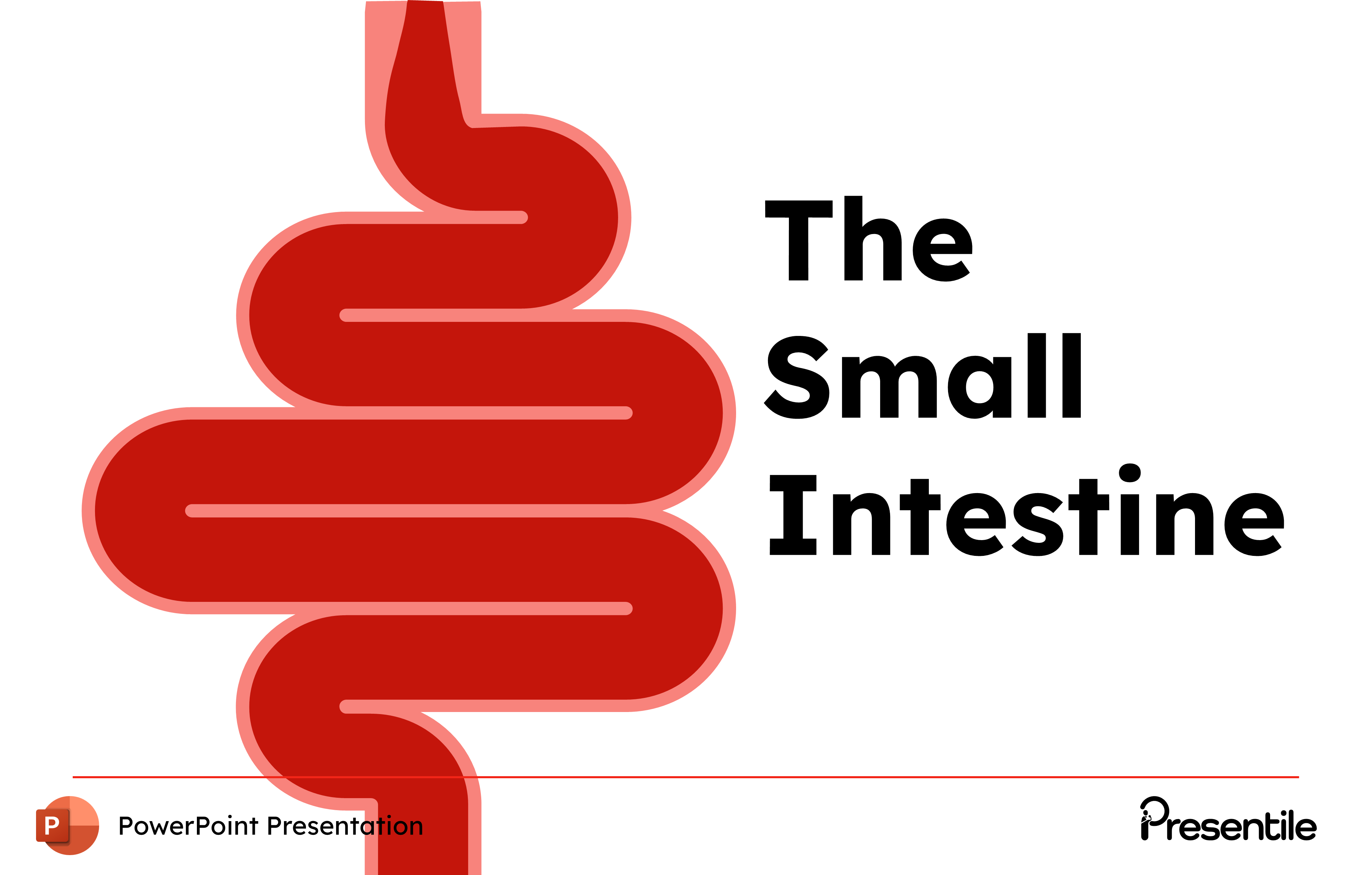
Slide 32: The Small Intestine Definition

The small intestine is A long tube-like organ that connects the stomach and the large intestine. It is about 6 meter long and folds many times to fit inside the abdomen.
Slide 33: Structure and Function of Duodenum, Jejunum, and Ileum

The small intestine is divided into three distinct regions, each with unique structural and functional characteristics:
The first section is the duodenum which is located closest to the stomach, receiving partially digested food, bile, and pancreatic juices to start breakdown.
The middle section is the Jejunum which is the longest and most coiled portion, where much of the absorption of nutrients occurs.
the final section is the ileum in which the process of nutrient absorption continues and transfers the remaining undigested material to the large intestine.
Each section is specialized to maximize nutrient breakdown and absorption, ensuring efficient digestion.
Slide 34: Villi, Microvilli, and Epithelial Cells for Nutrient Absorption

In the small intestine. the villi are finger-like projections, that significantly increase the surface area for nutrient absorption. Upon closer examination, we find microvilli, which are microscopic, brush-like projections on the surface of the villi, collectively forming what is known as the “brush border". These microvilli are composed of specialized epithelial cells that are adept at absorbing nutrients. while goblet cells are interspersed among them, secreting mucus to protect and lubricate the intestinal lining. these features highlight the remarkable adaptations of the small intestine for optimal nutrient uptake.
Slide 35: Histological Features and Layers for Nutrient Absorption

The small intestine’s histological structure consists of four distinct layers, each playing a vital role in digestion and nutrient absorption.
the Mucosa: This innermost layer contains villi and microvilli, and Specialized cells. responsible for both absorption and secretion.
the Submucosa: Located beneath the mucosa, this layer houses blood vessels, lymphatics, and nerves that support nutrient transport and communication within the digestive system.
the Muscularis: Comprising of smooth muscle, it is essential for peristalsis.
the Serosa: The outermost layer, composed of epithelial and connective tissue, provides lubrication and protection for the small intestine.
Each of these layers contributes to the small intestine’s efficiency in processing and absorbing nutrients.
Slide 36: Structure and Function from the Ileocecal Junction to the Anus

The large intestine extends from the ileocecal junction—where it connects to the small intestine—down to the anus.
Slide 37: Structure, Length, and Location in the Abdominal Cavity and Pelvis

the length of The large intestine is approximately 1.5 meters long and spans the abdominal cavity and pelvic region. This length provides ample surface area for these essential functions, ensuring efficient water reabsorption and waste management.
Slide 38: Anatomy and Function of the Colon, Cecum, Rectum, and Anal Canal

The large intestine begins at the cecum, a pouch-like structure that receives material from the small intestine. From there, it continues through the ascending colon, which extends upward along the right side of the abdomen. followed by the transverse colon. that crosses horizontally to the left side. Next, the descending colon runs vertically downward along the left side near the spleen. leading to the sigmoid colon. an S-shaped segment that curves toward the rectum. The rectum then serves as a storage area for feces until elimination. which occurs through the anal canal. Each segment of the large intestine contributes to absorbing water, compacting waste, and preparing it for elimination.
Slide 39: Anatomy of the Appendix and Mesoappendix

The vermiform appendix, commonly referred to as the appendix, is a small, tube-like structure extending from the cecum on the lower right side of the abdomen. It is connected to the ileum by a fold of mesentery known as the mesoappendix, which supplies the appendix with blood and helps anchor it within the abdominal cavity.
Slide 40: Anatomy and Function of the Ileocecal Sphincter
The cecum is located at the junction of the small and large intestines. Within the cecum lies the ileocecal sphincter, a muscular valve that prevents the backwards flow of ingested material from the cecum into the ileum. This sphincter ensures one-way movement of digested food, promoting efficient processing within the large intestine and preventing contamination of the small intestine.
Slide 41: Anatomy of the Internal and External Anal Sphincters

At the end of the rectum is the anal canal, which contains two key muscular structures. The internal anal sphincter, an involuntary ring of smooth muscle, automatically keeps the canal closed, preventing the passage of feces until defecation. Meanwhile, the external anal sphincter, a voluntary muscle, provides additional control over the release of waste. Together, these sphincters regulate the passage of fecal matter and maintain continence, combining automatic and voluntary control in the process of elimination.
Slide 42: Histological Features of the Large Intestine

The large intestine’s histological structure comprises distinct layers. each with specialized cells and features. The openings of intestinal glands secrete mucus, lubricating the intestinal lining to facilitate the smooth movement of waste. Absorptive cells, forming most of the epithelium, are crucial for water and electrolyte absorption. Goblet cells, abundant throughout, produce a mucus layer that protects the epithelium from abrasion and chemical damage. Scattered lymphatic nodules contain immune cells that guard against pathogens in the digestive tract. Together, these features enable the large intestine to perform essential roles in absorption, protection, and immunity.
Slide 43: Digestive System Physiology Key Organs and Functions

After gaining an overview of the anatomy of the digestive system, the next section will transition to its physiology.
Slide 44: Mouth Physiology The First Step in Digestion

The digestive tract starts to work as soon as food enters the mouth
Slide 45: Understanding Taste Receptors

The mouth is equipped with taste receptors, or taste buds, which detect various flavors and help the body distinguish nutrients from potentially harmful substances. taste buds recognize five basic sensations. Salty, triggered by sodium ions commonly found in salts. Sweet, associated with sugars and certain carbohydrates. Umami, a savory taste linked to glutamates in proteins. Bitter, often signaling the presence of alkaloids or toxins. and Sour, which arises from acids found in fruits and fermented foods. These sensations play a crucial role in guiding dietary choices and identifying potential toxins.
Slide 46: The Role of Saliva in Digestion

Saliva plays a crucial role in the digestive process by performing several key functions. It lubricates the food, making it easier to swallow, and it also contains the enzyme amylase, which begins the breakdown of complex carbohydrates (such as starches) into simpler sugars like maltose.
Slide 47: Role of Lysozyme in Fighting Bacteria with Saliva

In addition to its role in digestion, saliva contains lysozyme, an enzyme with antibacterial properties. Lysozyme helps protect the mouth from infections by breaking down the cell walls of certain bacteria, thus reducing the risk of bacterial growth.
Slide 48: Chewing Process Breaking Down Food into Smaller Pieces for Digestion

The chewing process is a mechanical action that breaks down food into smaller, more manageable pieces. This process, known as mastication, is performed by the teeth, which grind and crush the food, increasing its surface area. This step is crucial for preparing the food for further digestion in the stomach and intestines.
Slide 49: Deglutition or Swallowing Stages

Deglutition, or swallowing, is a coordinated process that safely transports food from the mouth to the stomach in three stages. The Oral Phase is voluntary: during this phase the food formed into the bolus and pushed to the back of the throat. consciously choosing when to swallow. Next is the Pharyngeal Phase, which is involuntary. In this stage, the bolus moves through the pharynx into the esophagus, with the soft palate rising to block the nasal passages. and the epiglottis covering the airway to prevent food from entering the trachea. Finally, the Esophageal Phase also occurs involuntarily. as peristaltic contractions push the bolus down the esophagus toward the stomach. where the lower esophageal sphincter relaxes to let it enter. Together, these stages ensure food moves efficiently and safely from the mouth to the stomach.
Slide 50: Chemical Digestion Role of Parietal and Chief Cells in Gastric Mucosa

As food enters the stomach, chemical digestion starts with the help of secretions from two specialized cell types in the gastric mucosa. Parietal Cells secrete hydrochloric acid, which lowers the stomach’s pH to create an acidic environment. This acidity helps breaking down proteins, activates enzymes, and kills potential pathogens. Meanwhile, Chief Cells release pepsinogen, an inactive enzyme precursor. In the acidic environment, pepsinogen converts to pepsin, an active enzyme that breaks down proteins into smaller peptides. Together, these cells enable the stomach to initiate chemical breakdown, preparing food for further digestion in the small intestine.
Slide 51: Role of Mixing and Peristaltic Waves in Food Breakdown

In the stomach, mechanical digestion complements chemical processes to ensure thorough food breakdown. This is achieved through two main types of muscular movements. The First one is Mixing Waves, which are rhythmic contractions that liquefy food. transforming it into chyme. These waves blend food with digestive juices. maximizing exposure to enzymes for better digestion. Then, Peristaltic Waves follow to create wave-like contractions that move the chyme gradually toward the pylorus. This controlled movement allows the chyme to flow into the duodenum at an optimal rate.
Slide 52: The Role of Bile Salts in Lipid Digestion

The liver and gallbladder work together to produce and store bile. which plays a key role in the digestion of fats. bile salts help emulsify large lipid globules into smaller lipid droplets. This process significantly increases the surface area of the lipids.
Slide 53: Key Enzymes for Digestion and Nutrient Breakdown

The exocrine pancreas plays a crucial role in digestion, acting as the body's primary source of digestive enzymes. These enzymes break down food into its absorbable components. Trypsin breaks down proteins into smaller peptides and amino acids, essential for protein digestion. Chymotrypsin, another enzyme involved in protein digestion, works alongside trypsin to further break down proteins. Carboxypeptidase assists in the final breakdown of peptides into amino acids, ensuring complete protein digestion. Pancreatic Amylase breaks down complex carbohydrates into simple sugars like maltose, facilitating carbohydrate absorption. Finally, Lipase breaks down lipids into fatty acids and glycerol, essential for the digestion and absorption of dietary fats. Together, these enzymes from the exocrine pancreas ensure that proteins, carbohydrates, and fats are effectively broken down and prepared for absorption in the small intestine.
Slide 54: The Small Intestine Section Slide
After protein digestion in the stomach The Small Intestine receives the Chyme and other enzymes for further digestion and absorption.
Slide 55: Chemical Digestion in the Small Intestine

the Chemical digestion in the small intestine continues to break down food into absorbable nutrients. Lactase is an enzyme that breaks down lactose into glucose and galactose, which can then be absorbed into the bloodstream. Similarly, Sucrase breaks down sucrose into glucose and fructose, enabling the body to absorb these simple sugars. The microvilli in the small intestine, which form the brush border, it help increase the surface area for nutrient absorption, ensuring the efficient uptake of sugars, amino acids, and fatty acids into the bloodstream for energy and metabolism.
Slide 56: The Large Intestine Section Slide

The undigested food enters The large Intestine, for the final processing and preparing it for elimination.
Slide 57: Water, Fiber, and Dead Cell Absorption in The Large Intestine

The final phase of digestion occurs in the large intestine. Water reabsorption, whereby the large intestine absorbs water from the residual food matter.. Fermentation of fiber also occurs. as dietary fibers that were not digested in the small intestine undergo fermentation by gut microbiota. Furthermore, Dead epithelial cells from the intestinal mucosa and indigestible matter are excreted and ultimately eliminated from the body as part of the feces. This phase is vital for conserving water and preparing waste for excretion.
Slide 58: The Gut Microbiome Supporting Digestion, Immunity, and Vitamin Synthesis

The gut microbiome is integral to digestive health and overall well-being, performing several key functions. Fiber breakdown is one of its primary roles. as gut microbiota ferment dietary fibers that the human digestive system cannot process. producing short-chain fatty acids. that promote colon health and provide energy. Additionally, certain gut bacteria are responsible for the synthesis of vitamin K, an essential nutrient for blood clotting and bone health. The microbiome also plays a critical role in immune system support by synthesizing antiviral proteins and encouraging the production of immune cells that help protect against pathogens. This interaction between the human host and its gut microbiota is essential for maintaining digestive, immune, and overall health.
Slide 59: Mass Movements and Haustral Churning in the Large Intestine

Mechanical Digestion and Motility in the Large Intestine involve several key processes that help move and compact the waste material:
Mass Movements: These are strong, peristalsis-like contractions that occur in the colon, propelling the stool towards the rectum. typically occur a few times a day and are responsible for moving the contents of the large intestine toward the distal colon for eventual elimination.
Haustral Churning: In addition to mass movements, the large intestine experiences slower contractions. These contractions mix the contents of the colon, facilitating the absorption of water and electrolytes.
Slide 60: The Role of Stretch Receptors and Parasympathetic Nerves

The defecation reflex. It begins, when stretch receptors in the walls of the large intestine, detect the stretching of the colon as it fills with feces. sending neural signals to the spinal cord. These signals are relayed via parasympathetic nerves to the descending colon, rectum, and anus, triggering the reflex.
Slide 61: Defecation Process Step-by-Step Breakdown of Stool Elimination

There is a series of coordinated steps that enable the elimination of waste from the body. beginning with. the relaxation of the internal anal sphincter, an involuntary response triggered by stretch receptors in the rectum signaling the brain. This allows stool to move closer to the anus. Following this, the external anal sphincter, which is under voluntary control, relaxes in response to brain signals, enabling further stool movement. Simultaneously, contraction of the abdominal muscles increases intra-abdominal pressure, assisting in pushing the stool out of the rectum. Finally, with both sphincters relaxed and sufficient pressure applied, the stool is expelled, completing the defecation process.
Slide 62: Thank you Ending Slide

This is an ending slide for the digestive system physiology and anatomy presentation
Features of
Digestive System Physiology & Anatomy PowerPoint Presentation
- Fully editable in PowerPoint
- All graphics are in vector format
- Medically Referenced information and data
 Slides count:
 Compatible with:Microsoft PowerPoint
 File type:PPTX
 Dimensions:16:9
- Non-animated PowerPoint
- Animated PowerPoint File
- Animated PowerPoint with Voice Over
- PDF Documents with presentation script


