


No items found.
Content of
Pneumothorax PowerPoint Infographics
Infographic 1: Understanding pneumothorax-collapsed lungs

- This infographic explains pneumothorax, also known as a collapsed lung.
- It defines the condition as air entering the pleural space, disrupting normal lung function.
- Key risk factors include trauma, medical procedures, and pressure injuries.
- Symptoms include sharp chest pain, rapid breathing, reduced chest expansion, and a hyper-resonant chest sound.
- The source references the American Thoracic Society and StatPearls for more information.
Infographic 2: Pneumothorax–Overview

- This infographic provides an overview of pneumothorax, describing it as air in the pleural space causing lung collapse.
- It categorizes causes into three types: primary (e.g., smoking, Marfan syndrome, pregnancy, tall-thin body), secondary (e.g., COPD, asthma, HIV with pneumocystis), and iatrogenic (e.g., medical procedures, Positive pressure ventilation).
- The diagnosis involves clinical evaluation, trauma history, and auscultation, while treatment includes oxygen therapy, chest tubes, and surgical interventions.
Infographic 3: Understanding pneumothorax–clinical signs & differential diagnosis

- This Infographic is about understanding Pneumothorax, it presents its clinical signs such as sudden chest pain, cyanosis, tachycardia, unequal chest rise and fall, low blood pressure, and deviated trachea.
- Differential diagnoses include pleural effusion, where fluid in the pleural space causes lung collapse; hemothorax, resulting from blood in the pleural space; pneumothorax, where air causes the collapse; and tension pneumothorax, a critical condition where trapped air in the pleural space worsens the collapse and can lead to severe complications if untreated.
Infographic 4: Understanding diagnostic methods and signs of pneumothorax

- This infographic explains pneumothorax, diagnosis methods, and different types.
- The top section defines pneumothorax, three diagnostic approaches are shown: lung auscultation, laboratory tests (ABG analysis), and imaging via chest radiography.
- The bottom section details Signs according to Types : spontaneous (no clinical signs initially), tension (causing hypotension and hypoxia), iatrogenic (due to medical procedures), and catamenial (affecting women during menstruation).
- The design uses a clean, medical-themed color scheme of light blue, pink, and dark teal, with clear icons and illustrations to convey information.
Infographic 5: Smoking as a risk factor of pneumothorax

- The infographic emphasizes that cigarette smoking significantly increases the risk of developing a first pneumothorax, with additional risk factors including weight (based on height-weight ratio) and pregnancy (which requires special consideration for diagnosis when chest pain and dyspnea are present).
- The condition shows distinct age-related patterns, with primary spontaneous pneumothorax typically occurring in younger adults aged 20-30 years.
- Secondary spontaneous pneumothorax is more common in older adults aged 60-65 years. Regarding prognosis, PSP (Primary Spontaneous Pneumothorax) is generally benign and often resolves without major intervention, though it can recur for up to three years.
- The recurrence rates differ between types, with PSP having a 30% recurrence rate and SSP (Secondary Spontaneous Pneumothorax) having a higher rate of 43% over five years.
Infographic 6: Pneumothorax incidence & gender disparity

- This infographic detailing the incidence of pneumothorax showing a significant gender disparity, with men experiencing rates of 7.4-18 per 100,000 compared to women at 1.2-6 per 100,000, representing a male-to-female ratio of 3:1.
- Primary Spontaneous Pneumothorax (PSP) predominantly affects young adults aged 20-30 years, with peak incidence in the early 20s.
- The infographic reveals that up to 10% of patients may be asymptomatic, and some with mild symptoms might not seek medical care.
- The main risk factors for developing pneumothorax are identified as genetic predisposition, smoking, underlying lung disease, and sporadic occurrence.
Infographic 7: Pneumothorax diagnosis & symptoms

- This Pneumothorax infographic showcasing the symptoms: chest pain, cough, shortness of breath, cyanosis, rapid heart rate, and fast breathing.
- The infographic emphasizes that anyone experiencing symptoms of a collapsed lung should seek immediate emergency care.
- For diagnosis, healthcare providers use two main approaches: physical examination and imaging techniques.
- The imaging options include chest X-ray as the primary diagnostic tool, CT scan for complex cases, and ultrasound for emergency settings.
Infographic 8: Pneumothorax at a glance

- The infographic emphasizes that pneumothorax is an urgent medical condition requiring immediate treatment, illustrated with a clear diagram of the lungs showing air in the pleural space.
- It highlights two key risk factors: male gender (men being more susceptible than women) and smoking.
- The treatment section outlines four main options: supplemental oxygen administered through a nasal canula at 3 L/min, one-way valve insertion (a portable system to avoid hospital admission), chest tube placement into the pleural space, and thoracostomy lung surgery which is noted as safe for most patients.
Infographic 9: Pneumothorax a simplified overview infographic

- The prevalence data shows a significant gender disparity, with males having a rate of 6.3 per 100,000 compared to females at 2 per 100,000, resulting in a 3:1 male-to-female ratio.
- The infographic identifies three major complications: pain and skin infection at the tube thoracostomy site, respiratory failure due to inadequate oxygenation, and cardiac arrest which occurs specifically with tension pneumothorax.
- There are three main types of pneumothorax presented: tension pneumothorax (where air enters the pleural space through the chest wall or airways), spontaneous pneumothorax (caused by rupture of a bleb on the lung surface), and traumatic pneumothorax (resulting from trauma to the chest wall or pleural membrane disruption).
Infographic 10: Pneumothorax risk factors, complications, and symptoms

- This infographic provides an overview of pneumothorax.
- The risk factors include family history, pregnancy, having a tall and thin body type, and Marfan syndrome.
- The main symptoms, include chest pain, bluish skin, dry cough, and an increased heart rate.
- The center of the image shows a person experiencing breathing difficulty, with an illustration of affected lungs.
- Severe complications, can include pulmonary edema, infection, respiratory failure, and heart failure.
Infographic 11: Pneumothorax examination findings & risk factors

- This Infographic details the risk factors and examination findings of pneumothorax, dividing risk factors into two categories: primary spontaneous (including tall thin body, Marfan Syndrome, and familial pneumothorax) and secondary spontaneous (including cyst fibroses, bronchogenic carcinoma, and thoracic endometriosis).
- Examination findings include respiratory discomfort, increased respiratory rate, and asymmetrical lung expansion, while tension pneumothorax can lead to more severe symptoms like tachycardia, respiratory failure, and cardiac arrest.
- The condition has a significant gender disparity in occurrence rates, affecting 7 per 100,000 males compared to 1 per 100,000 females.
- Pneumothorax may be challenging to diagnose through physical examination alone, suggesting the need for additional diagnostic methods.
Infographic 12: Primary spontaneous pneumothorax

- This infographic details Primary Spontaneous Pneumothorax (PSP), defining it as the spontaneous presence of air in the pleural space without underlying lung disease.
- The condition shows a significant gender disparity, and has a concerning recurrence rate of 20-60% worldwide.
- The main symptoms include shortness of breath, chest pain, and tachycardia, while potentially confusing conditions that could present similarly include embolism, pleuritis, and pneumonia.
- The image indicates that diagnosis can be confirmed through various imaging methods including CT scan, ultrasonography, and radiographs, emphasizing the importance of proper diagnostic testing for accurate identification.
Infographic 13: Pneumothorax collapsed lung- breathless moments

- This Infographic provides a comprehensive overview of pneumothorax, highlighting its key aspects in five main sections.
- The anatomical illustration shows air in the pleural space, which defines pneumothorax as a collapsed lung condition.
- The demographic data indicates that men are three times more likely to be affected than women, and the differential diagnoses include myocardial infarction and pulmonary embolism.
- Patient education focuses on three key restrictions: no diving, no smoking, and avoiding air travel.
- The treatment options range from oxygen therapy and needle aspiration to more invasive procedures like chest tube insertion and lung surgery.
Infographic 14: A comprehensive guide to pneumothorax

- This infographic presents a comprehensive guide to pneumothorax, explaining it as a condition.
- The guide emphasizes the serious nature of the condition, stating that some cases can be life-threatening and require medical monitoring.
- Four main causes are identified: chest injury, lung disease, ruptured air blisters on the lungs, and mechanical ventilation.
- The guide also outlines three key risk factors - smoking, genetic predisposition , and previous pneumothorax.
Infographic 15: A deep dive into pneumothorax

- This infographic provides a deep dive into pneumothorax.
- The condition is defined, primarily affecting patients between 18 and 40 years of age, with a significant male-to-female ratio of 5:1.
- Clinical findings include respiratory discomfort, decreased tactile fremitus, increased respiratory rate, hyperresonant percussion note, asymmetrical lung expansion, and decreased intensity of breath sounds.
- The condition has a concerning recurrence rate of 25-50%, with most recurrences happening within the first year.
- The differential diagnosis includes several serious conditions that need to be ruled out: aspiration, bacterial or viral pneumonia, acute aortic dissection, myocardial infarction, and diaphragmatic injuries.
- The image effectively shows that pneumothorax is a condition requiring careful medical attention and precise diagnosis due to its potential severity and similarity to other serious conditions.
Infographic 16: Insights into pneumothorax

- This infographic provides insights into pneumothorax, detailing several key aspects of the condition.
- The incidence rates show a significant gender disparity, and there's a 30% chance of recurrence within 3 years for primary pneumothorax.
- The infographic identifies five major risk factors: smoking history, existing lung conditions, tall and slim body type, family history of pneumothorax, and inflammation in small airways.
- Serious complications can include respiratory failure, pulmonary edema, pneumohemothorax, pneumopericardium, and pneumoperitoneum, with the image emphasizing the critical importance of seeking immediate emergency medical attention when symptoms appear.
Infographic 17: Pneumothorax-answering important questions

- This infographic explains the pneumothorax by answering three questions what is pneumothorax?
- Difference between an open and closed pneumothorax?
- Types of pneumothoraxes.
Infographic 18: Pneumothorax explained

- Defining pneumothorax, and bilateral pneumothorax is when air leaks out of both lungs and causes them to collapse, with the recurrence rates and the causes including mechanical ventilation, lung disease, and ruptured air blisters.
- Risk factors of the pneumothorax are smoking, genetics, and previous pneumothorax.
Infographic 19: Breaking down pneumothorax

- Breaking down pneumothorax infographic include: Diseases associated with secondary spontaneous pneumothorax like COPD, Asthma, HIV, Tuberculosis, cystic fibrosis.
- The recurrence rates of pneumothorax 25% to 50%, with ratio male to female 3:1.
- Diagnosis is based on clinical criteria and chest x-ray.
- Most pneumothoraxes require transcatheter aspiration or tube thoracostomy.
- Treatment depends on the type, size, and effects of the pneumothorax.
Infographic 20: Collapsed lung ( pneumothorax) treatment & diagnosis

- This infographic outlines the treatment and diagnosis procedures for pneumothorax: The treatment section presents four main approaches in order of increasing intervention: observation, needle aspiration or chest tube insertion, nonsurgical repair (when chest tubes don't re-expand the lung), and surgery.
- The diagnosis process begins with a healthcare provider taking a medical history and performing a physical exam, with particular attention to symptoms in cases of tension pneumothorax.
- For imaging-based diagnosis, three options are available: chest X-ray as the primary diagnostic tool, CT scan for more detailed imaging when needed, and ultrasound for identifying pneumothorax.
Infographic 21: Pleural space air leakage pneumothorax explained

- This infographic provides a comprehensive explanation of pneumothorax and its various aspects.
- What is pneumothorax? incidence rates of pneumothorax.
- Secondary pneumothorax can be caused by several underlying conditions: COPD, asthma, and cystic fibrosis.
- The main risk factors are identified as smoking, tall and thin build, and male sex.
- The mortality rate for secondary spontaneous pneumothorax is shown as 10%.
- Tension pneumothorax can develop through three main pathways: penetrating/blunt trauma, mechanical ventilation, or conversion from a simple pneumothorax to tension pneumothorax.
Infographic 22: Pneumothorax signs

- Pneumothorax occurs when air leaks into the space between one of the lungs and the chest wall, and it disproportionately affects males more than females, with incidence rates ranging from 7.4 to 37 per 100,000 per year for males and 1.2 to 15.4 per 100,000 per year for females.
- The primary signs and symptoms of pneumothorax include sharp, stabbing chest pain, shortness of breath, a dry hacking cough, rapid breathing and heartbeat, cyanosis, and low blood pressure.
- Medical professionals strongly emphasize that immediate medical attention should be sought if someone experiences sudden chest pain, shortness of breath, rapid breathing, or any bluish discoloration of the skin.
Infographic 23: Pneumothorax pathophysiology & risk factors

- Pneumothorax can develop from chest trauma, excess lung pressure, or lung disease.
- The pathophysiology involves air entering the pleural space either from outside the chest or from the lung itself through mediastinal tissue planes or direct pleural perforation, resulting in increased intrapleural pressure and decreased lung volume.
- There are three main classifications: primary spontaneous pneumothorax, secondary spontaneous pneumothorax, and traumatic pneumothorax. risk factors, including smoking, genetic predisposition, and having a previous pneumothorax.
Infographic 24: Approach to pneumothorax

- This infographic outlines the approach to pneumothorax, with its definition.
- The three diseases most associated with pneumothorax are COPD, asthma, and cystic fibrosis.
- The treatment options presented include needle aspiration (where a hollow needle with a small flexible catheter is inserted between the ribs into the air-filled space), chest tube insertion (involving the placement of a flexible tube into the air-filled space), and surgery (which can be performed through small incisions using a tiny fiber-optic camera and narrow, long-handled surgical tools).
- Epidemiologically, the infographic notes that primary spontaneous pneumothorax occurs more frequently in men than in women.
Infographic 25: A short review of Pneumothorax

- This infographic provides a short review of pneumothorax, defined as the presence of air in the pleural cavity.
- The age distribution indicates that pneumothorax peaks in individuals aged 16-64 years, with a pediatric incidence of 5-10 cases per 100,000 children under 18 years.
- The complications shown include failure of the lung to expand, re-expansion pulmonary edema, and air leaks.
- Common signs and symptoms include dyspnea, tachycardia, tachypnea, and hypotension. The diagnostic investigations typically involve chest ultrasound, CT chest, and arterial blood gas analysis. Medical conditions that can cause pneumothorax include asthma, pneumonia, COPD, and cystic fibrosis.
Infographic 26: A review of Pneumothorax

- This infographic is about a review of pneumothorax, characterized by the presence of air in the pleural space causing partial or complete lung collapse, shows a notable gender disparity.
- Several risky conditions can lead to this condition, including lung diseases, injuries that can puncture the lung, and mechanical ventilation.
- Treatment approaches are guided by several factors: gender; body type; and smoking habits.
Infographic 27: Pneumothorax a brief summary

- This Infographic provides a brief summary Pneumothorax, shows significant gender differences in prevalence.
- The condition can be diagnosed through several imaging methods: chest X-rays in some cases, CT scans, and ultrasound imaging.
- The main causes include chest injury, lung disease, and ruptured air blisters.
- Serious complications can arise from pneumothorax, including respiratory failure, cardiac arrest, and empyema.
Infographic 28 : Pneumothorax an overview definition, warning signs, and differential diagnosis

- This infographic includes the pneumothorax definition as well as warning signs including chest pain on one side, shortness of breath, and cyanosis (where skin, lips, and nails take on a bluish tone).
- Diagnosing pneumothorax, several other conditions must be considered in the differential diagnosis: diaphragmatic injuries (often associated with thoracic and abdominal organ injuries), pulmonary embolism (a sudden blockage in pulmonary arteries), myocardial infarction (when blood flow to the heart muscle is suddenly cut off), acute pericarditis (inflammation of the pericardium), and aspiration pneumonia (a lung infection caused by inhaled oral or gastric contents).
Infographic 29 : Pneumothorax a brief overview severe symptoms & common causes

- Pneumothorax a brief overview.
- The condition presents symptoms including easy fatigue, chest tightness, shock and collapse, and abnormal breathing patterns.
- There are multiple common causes: injury to the lung, air blisters of the lung (blebs), air pressure changes (from activities like scuba diving or high-altitude travel), tall and thin people and smokers (due to higher frequency of blebs in the apices), mechanical ventilation (which can create an imbalance of air pressure within the chest), and lung diseases (such as COPD, cystic fibrosis, lung cancer, or pneumonia).
- The infographic emphasizes that immediate emergency care should be sought if chest pain becomes severe or breathing becomes increasingly difficult.
Infographic 30 : Pneumothorax a quick review common symptoms, causes, and prevention

- This Infographic provides a quick review of pneumothorax, which occurs when air accumulates in the pleural space of the lungs.
- The condition has three main causes: chest trauma, excess pressure on the lungs, and underlying lung disease.
- Common symptoms presented include bluish skin coloration, shoulder pain, and nasal flaring. Regarding prevention, while there is no known way to completely prevent a collapsed lung, risk can be reduced by following proper scuba diving procedures and avoiding smoking.
Infographic 31 : What is Pneumothorax?

- This Infographic answers what is Pneumothorax , which is defined as an abnormal accumulation of air or gases in the pleural space around the lungs, requiring immediate treatment upon diagnosis.
- The Infographic outlines three distinct types: Primary Spontaneous, Tension, and Traumatic.
- The vital signs to monitor include tachycardia, tachypnea, hypoxia, and hypotension.
- The infographic emphasizes the medical urgency of this condition, reinforcing the importance of proper diagnosis.
Infographic 32 : Pneumothorax possible treatment & Prognosis

- This infographic provides information about pneumothorax, also known as a collapsed lung.
- The infographic explains how to measure the severity of a pneumothorax using chest radiographs, where measurements less than 3 cm from the apex indicate a small pneumothorax, while those greater than 3 cm indicate a large one.
- The treatment options presented include chest drain insertion, pleurodesis which involves adhering the lung to the chest cavity, and various surgical interventions.
- The infographic also includes a notable prognosis detail, stating that while up to 50% of pneumothorax patients may experience a recurrence, there are typically no long-term complications after successful treatment.
Infographic 33 : Pneumothorax disease background

- This infographic provides a disease background of pneumothorax, explaining that it occurrence.
- The etiology is categorized into three types: primary, secondary, and traumatic.
- The history section outlines three main symptoms that patients typically experience: sudden chest pain, shortness of breath, and fast heart rate.
- Three serious complications that can arise: pulmonary edema following treatment, pneumopericardium, and pneumohemothorax.
Infographic 34: Pneumothorax primary spontaneous vs secondary spontaneous

- This infographic compares primary and secondary spontaneous pneumothorax (PSP and SSP), highlighting their key differences and characteristics.
- Primary spontaneous pneumothorax occurs due to spontaneous rupture of subpleural blebs or bullae, often linked to smoking or inherited factors, while secondary spontaneous pneumothorax typically affects patients with severe chronic obstructive pulmonary disease.
- The common symptoms for both types include tachycardia, hypotension, and cyanosis, while lifestyle factors that can contribute to the condition include inhaled drugs, smoking, and deep-sea diving.
- The prognosis section reveals a notable difference in outcomes: PSP has a 30% recurrence rate over five years and isn't considered a significant health threat, whereas SSP has a higher 43% recurrence rate and is more lethal due to underlying lung disease and pneumothorax size.
Infographic 35 : Pneumothorax primary spontaneous in USA
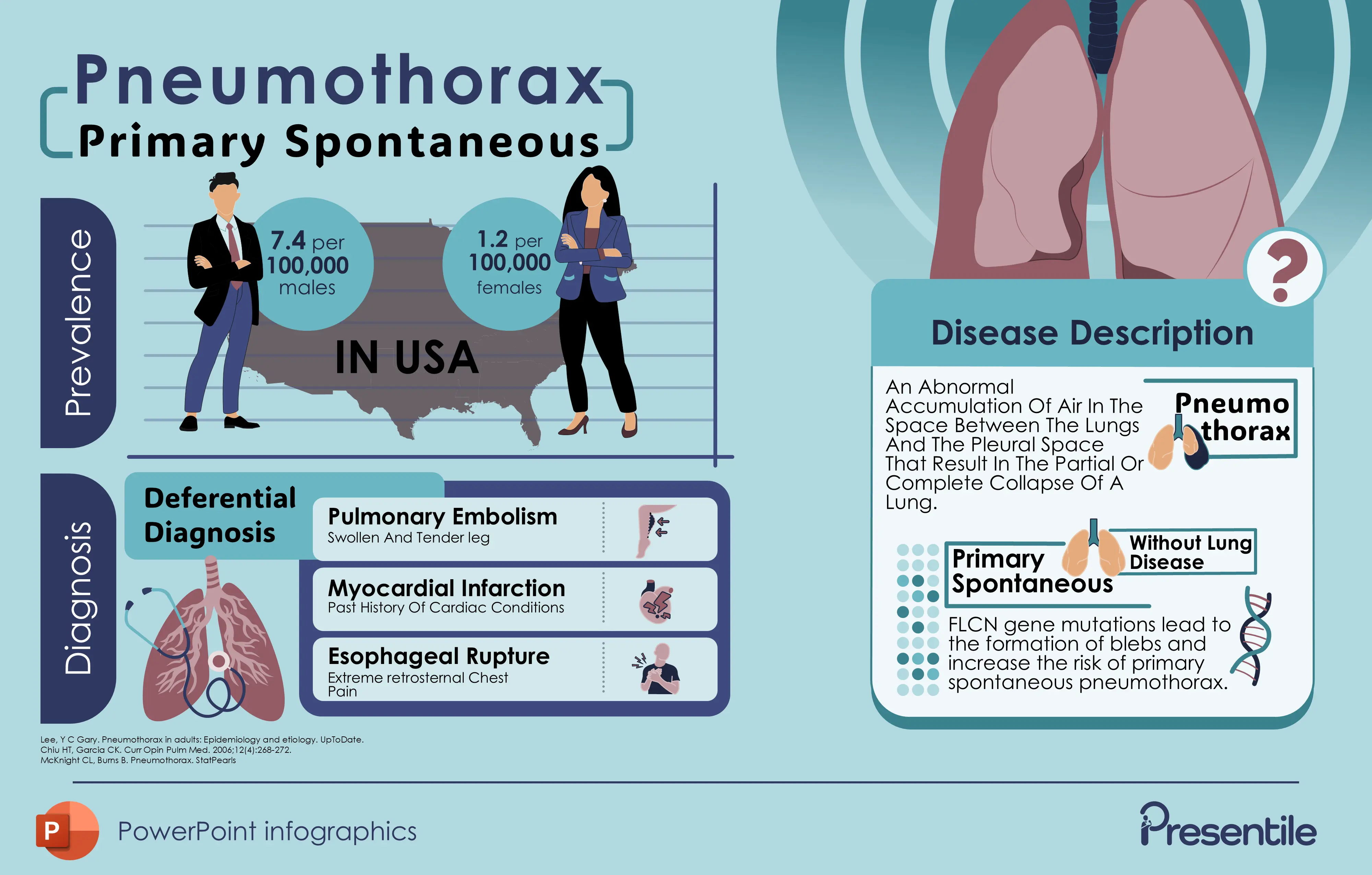
- This infographic provides detailed information about Primary Spontaneous Pneumothorax (PSP), including its prevalence, diagnosis, and disease description.
- In the USA, there is a notable gender disparity in prevalence, with 7.4 cases per 100,000 males compared to 1.2 cases per 100,000 females.
- The differential diagnosis section identifies three conditions that should be considered when diagnosing PSP: pulmonary embolism (characterized by swollen and tender leg), myocardial infarction (associated with past cardiac conditions), and esophageal rupture (marked by extreme retrosternal chest pain).
- The disease description explains that pneumothorax is an abnormal accumulation of air between the lungs and pleural space, which can cause partial or complete lung collapse, and notably mentions that FLCN gene mutations can lead to the formation of blebs and increase the risk of primary spontaneous pneumothorax.
Infographic 36: Pneumothorax forms, gender disparities

- This infographic provides detailed information about different aspects of pneumothorax and its manifestations.
- The infographic explains that pneumothorax can be divided into two forms: open pneumothorax, where air enters from outside due to injury, and closed pneumothorax, where air enters from inside due to overpressure.
- There's a significant gender disparity in Secondary Spontaneous Pneumothorax, with a male-to-female ratio of 3:1.
- The infographic details that severe cases may present with three main symptoms: cough or palpitations, lightheadedness, and sharp pain on the lateral side of the body localizing to one side.
- Finally, it notes that pneumothorax presents differently in infants, with distinctive signs including cyanosis, tachypnea, and decreased breath sounds.
Infographic 37: Pneumothorax case history, investigations, types, and risk factors

- This infographic provides a simple overview pneumothorax, The case history section shows that patients are typically present with dyspnea and pleuritic chest pain, with diagnosis confirmed through chest X-rays that show a gap between the lung and chest wall.
- There are three main types: primary pneumothorax, secondary pneumothorax, and iatrogenic pneumothorax.
- Key risk factors highlighted in the image include smoking, and genetics, as certain types tend to run in families.
Infographic 38: Diagnosis, and common symptoms pneumothorax

- This infographic details additional aspects of pneumothorax, highlighting that it occurs 3 to 6 times more frequently in males than females.
- The condition's common symptoms include pleuritic chest pain, reduced chest expansion, and reduced breath sounds on the affected side, with the important note that these symptoms can mimic other serious conditions like pericarditis, pneumonia, pleuritis, and pulmonary embolism.
- For diagnosis, doctors rely on clinical diagnosis, auscultation, X-rays, and assessment of any present trauma.
- Notably, the recurrence rate is significant, ranging from 25% to 50%, with the highest risk of recurrence occurring within the first 30 days after the initial episode.
Infographic 39: Diagnosis, and common symptoms pneumothorax

- This infographic details additional aspects of pneumothorax, highlighting that it occurs 3 to 6 times more frequently in males than females.
- The condition's common symptoms include pleuritic chest pain, reduced chest expansion, and reduced breath sounds on the affected side, with the important note that these symptoms can mimic other serious conditions like pericarditis, pneumonia, pleuritis, and pulmonary embolism.
- For diagnosis, doctors rely on clinical diagnosis, auscultation, X-rays, and assessment of any present trauma.
- Notably, the recurrence rate is significant, ranging from 25% to 50%, with the highest risk of recurrence occurring within the first 30 days after the initial episode.
Infographic 40: How to diagnose pneumothorax & key points

- This infographic provides detailed information about diagnosing pneumothorax and its key characteristics.
- The diagnostic process involves two main steps: lung ultrasound (which looks for absence of lung sliding), and chest X-ray (which shows a visible rim between the lung margin and chest wall).
- The image highlights two main types of spontaneous pneumothorax: primary, which occurs in tall, thin young men in their teens and 20s without underlying disease, and secondary, which happens in patients with existing pulmonary disease, often due to rupture of a bleb or bulla in those with severe chronic obstructive pulmonary disease.
Infographic 41: The types of pneumothorax & risk factors

- This infographic outlines the four types of pneumothorax and their risk factors.
- The types include primary spontaneous, secondary spontaneous, iatrogenic, and traumatic.
- The risk factors are divided into two categories: traumatic (including contact sports, chest-related medical procedures, and assisted respiratory care) and nontraumatic (including family history, smoking history, and existing lung conditions like asthma or COPD).
Infographic 42: Reasons behind a collapsed lung (pneumothorax) symptoms and treatment

- This infographic provides reasons, describing it as a collection of air in the pleural space with three main types: primary, secondary, and tension.
- The symptoms section outlines that patients usually suffer from shortness of breath, bluish skin/nails, cough, fast heart rate, fast breathing, and fatigue - with an urgent warning to seek immediate emergency care if these symptoms appear.
- Three main causes of collapsed lungs are identified: chest injury, lung diseases, and mechanical ventilation.
- The treatment section specifically mentions tube thoracostomy as a treatment method for secondary and traumatic pneumothoraxes.
Infographic 43: Who is in risk for pneumothorax etiology & types

- This infographic provides a detailed breakdown of pneumothorax risk factors, causes, and classifications.
- For risk factors, it identifies four main groups: smokers, those with genetic predisposition, individuals with previous pneumothorax, and notably mentions that men are generally at higher risk than women.
- The etiology section lists four medical conditions that can cause pneumothorax: asthma, foreign body aspiration, malignancy, and pulmonary abscess.
- The bottom of the image divides pneumothorax into two main categories - Primary Spontaneous and Secondary Spontaneous, all centered around the core concept of air presence in the pleural space.
Infographic 44: Pneumothorax clinical presentation

- This infographic details the clinical presentation of pneumothorax, noting that the incidence of non-traumatic cases is 7.4-18 per 100,000 people per year.
- The infographic emphasizes the importance of watching for signs of tension pneumothorax, including tracheal deviation, sudden difficulty ventilating, hypotension, or distended neck veins.
- The main clinical presentations in adults include sudden sharp pain on the lateral side of the body, lightheadedness with dizziness and fainting feeling, complaints of cough or palpitation, and chest pain that varies according to type.
- For infants, the presentation is notably different, with four distinct signs: cyanosis, nasal flaring, tachypnea, and decreased breath sounds, indicating how the condition manifests differently in this age group.
Infographic 45: The pharmacotherapy of pneumothorax & complications

- This infographic details treatment approaches and complications of pneumothorax, which is defined as an accumulation of air or gas in the pleural space.
- The pharmacotherapy section explains that treatment focuses on pain management, both from the condition itself and from procedures like thoracostomy or needle aspiration, utilizing local anesthetics at the thoracostomy site and intravenous or oral pain medications.
- The complications section identifies four major potential issues: conversion to tension pneumothorax, re-expansion pulmonary edema, hypoxemic respiratory failure, and iatrogenic complications that can arise from needle decompression or thoracostomy procedures.
- The central illustration effectively shows the anatomical impact of pneumothorax on the lungs, with arrows indicating the direction of air pressure.
Infographic 46: How to manage pneumothorax?

- This infographic provides a management plan for pneumothorax, a condition where air enters the pleural space and causes lung collapse.
- The diagnostic process involves three key investigations: CXR (chest X-ray) to confirm clinical suspicion, ABG (arterial blood gas) which may be normal in acute cases, and ECG to rule out cardiac causes.
- For management, the image outlines two distinct approaches: observation alone is sufficient for asymptomatic patients with small pneumothorax (<3cm or <20% of hemithorax), while tube thoracostomy is necessary for larger primary spontaneous pneumothorax or tension pneumothorax.
- Finally, the prognosis section warns that spontaneous pneumothorax has a significant recurrence rate of 20-60% within three years of the initial episode.
Infographic 47: History & management of pneumothorax

- This infographic outlines the key aspects of pneumothorax, starting with its definition.
- The history section identifies three main presentations: pleuritic chest pain, underlying conditions like COPD and cystic fibrosis, and cases that are either spontaneous or related to medical procedures/trauma.
- The management approach is divided into two pathways: simple observation for small primary pneumothorax without significant breathing difficulties, while patients with more severe symptoms or secondary pneumothorax require more intensive intervention including supportive oxygen therapy and either needle aspiration or chest drain placement.
- The anatomical illustration on the right shows how air in the pleural space (indicated by red arrows) can affect the lungs.
Features of
Pneumothorax PowerPoint Infographics
- Fully editable in PowerPoint
- All graphics are in vector format
- Medically Referenced information and data
Pneumothorax PowerPoint Infographics
Pneumothorax PowerPoint Infographics
Price:
$ 29.00 USD
Specifications
 Slides count:
 Compatible with:Microsoft PowerPoint
 File type:PPTX
 Dimensions:16:9
Files Included
- Non-animated PowerPoint
- Animated PowerPoint File
- Animated PowerPoint with Voice Over
- PDF Documents with presentation script
Elevate Your Work with Our Innovative Slides
Thank you! Your submission has been received!
Oops! Something went wrong while submitting the form.


